How To Measure Scoliosis On An X-ray? What Your Xrays Mean

When it comes to how to measure scoliosis on an X-ray, this involves determining a patient’s Cobb angle measurement and angle of trunk rotation (ATR), and it’s important that these measurements are accurate because treatment plans are shaped around them.
When it comes to diagnosing scoliosis, an X-ray is needed to confirm what’s happening in and around the spine, and as a minimum Cobb angle measurement of 10 degrees is the condition’s diagnostic cutting-off point, an X-ray is also needed to officially diagnose scoliosis.
The first use of an X-ray for scoliosis is in diagnosing the condition, so let’s start there.
Table of Contents
Diagnosing Scoliosis
Current estimates of the Scoliosis Research Society have close to seven million people currently living with scoliosis in the United States alone, and it is the leading spinal condition amongst school-aged children.
Scoliosis is so prevalent there was a time when mandatory scoliosis screening was performed in schools across the States, but that has since changed, shifting the onus of recognizing the condition’s early signs to parents and patients themselves.
Scoliosis can’t be treated until it’s diagnosed, and diagnosing scoliosis isn’t always easy, particularly when mild, and this is where an X-ray comes in.
Scoliosis involves the development of an unnatural sideways spinal curve that also rotates, making it a complex 3-dimensional condition.
And in addition to the rotational component, an unnatural lateral spinal curve also has to be of a minimum size to be officially diagnosed as scoliosis: minimum Cobb angle measurement of 10 degrees.
A patient’s Cobb angle is known as the gold standard in the diagnosis and assessment of scoliosis, and this is because it shapes the crafting of effective treatment plans.
Cobb Angle and X-Ray
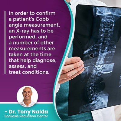
 In order to confirm a patient’s Cobb angle measurement, an X-ray has to be performed, and a number of other measurements are taken at the time that help diagnose and assess conditions.
A patient’s Cobb angle is determined by drawing lines from the tops and bottoms of the curve’s most-tilted vertebrae, and the resulting angle is expressed in degrees.
In a healthy spine with its healthy curves and alignment in place, the spine’s vertebrae (bones) are stacked on top of one another in a straight and neutral position, but if one or more vertebral bodies become unnaturally tilted, this shifts their position and can force the spine out of alignment.
The higher a Cobb angle, the further out of alignment the spine is, and the more severe the condition:
- Mild scoliosis: Cobb angle measurement of between 10 and 25 degrees
- Moderate scoliosis: Cobb angle measurement of between 25 and 40 degrees
- Severe scoliosis: Cobb angle measurement of 40+ degrees
- Very-severe scoliosis: Cobb angle measurement of 80+ degrees
So if condition indicators are detected in a screening exam, the next step is an X-ray to truly see what’s happening in and around the spine, to confirm a patient’s Cobb angle measurement, and to officially reach a diagnosis of scoliosis.
So what else does a scoliosis X-ray tell us?
What Else Does a Scoliosis X-Ray Measure and Tell Us?
The complex nature of scoliosis necessitates the customization of effective treatment plans, and this is because no two cases of scoliosis are the same, and treatment plans are shaped around the results of scoliosis X-rays.
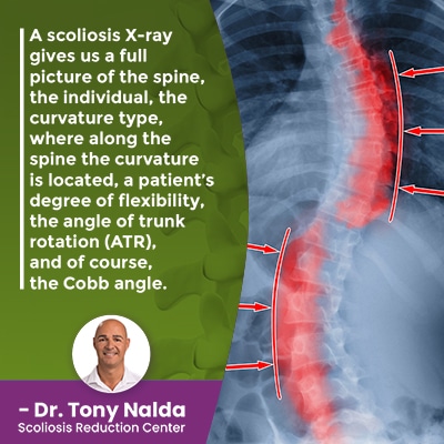
A scoliosis X-ray gives us a full picture of the spine, the individual, the curvature type, where along the spine the curvature is located, a patient’s degree of flexibility, the angle of trunk rotation (ATR), and of course, the Cobb angle.
Different curvature types mean different things, so this is an important part of further classifying conditions; for example, dextroscoliosis refers to typical conditions where the curve bends to the right, away from the heart, but levoscoliosis is atypical as the curve bends to the left, towards the heart.
When I see a scoliotic curve that bends to the left, this is a red flag that there is an underlying pathology at play, and this will change the course of treatment.
In addition, scoliosis can involve a single unnatural spinal curve, or the spine can curve in two opposite directions, classified as an S-curve or a double-curve scoliosis, and in these cases, an X-ray is needed to determine which is the major and minor curve as this shapes treatment.
There are three main spinal sections: the cervical spine (neck), thoracic spine (middle/upper back), and the lumbar spine (lower back).
Scoliosis can develop in any of the spine’s main sections, and can involve more than one section as a combined scoliosis; for example, thoracolumbar scoliosis involves a curve that engages the upper lumbar spine and the lower thoracic spine.
An X-ray is needed to classify the type of curve, where it’s located, and this tells me where to concentrate my treatment efforts.
A patient’s degree of flexibility is helpful to know because the more flexible a spine is, the more responsive it can be to treatment, and as scoliotic spine doesn’t just bend unnaturally to the side, but also twists, a patient’s angle of trunk (ATR) rotation is important because treatment that doesn’t address the rotational component of scoliosis is limited in its potential efficacy.
In addition, X-rays can be necessary throughout treatment to monitor for progression, and observe how the spine is responding to treatment so plans can be adjusted accordingly.
Monitoring for Scoliosis Progression
 Scoliosis is a progressive condition, meaning it has it in its nature to worsen over time, and this needs to be understood in order to be addressed effectively.
Where a scoliosis is at the time of diagnosis is not indicative of where it will stay, at least not without the efforts of proactive treatment that work towards counteracting the condition’s progressive nature.
As scoliosis progression is triggered by growth, children are most at risk for progression, and in the condition’s most-prevalent form, adolescent idiopathic scoliosis, diagnosed between the ages of 10 and 18, rapid-phase progression is possible due to the rapid and unpredictable growth spurts of puberty.
So when treating scoliosis in children, monitoring for progression is a focus of treatment, and this is done through periodic X-rays to re-assess and confirm a patient’s Cobb angle throughout treatment.
When it comes to treating adolescent idiopathic scoliosis, I will order X-rays with as little as an inch of growth because that growth can make the condition progress, so I need to see how growth affects progression to help determine a patient’s progressive rate, and I also want to see how patients are responding to treatment.
Part of the benefit of a conservative chiropractic-centered treatment approach is that it integrates multiple treatment disciplines into treatment plans so conditions can be impacted on every level, but this means knowing how to apportion these disciplines accordingly, and this is also determined by observing how the spine is responding to treatment.
When treating adolescent idiopathic scoliosis, the main treatment goal is to achieve a curvature reduction on a structural level, and then hold it there, despite the constant trigger of growth.
X-rays can also help estimate how much growth a patient has yet to go through: a key piece of information considering that growth is what triggers progression.
Conclusion
So when it comes to how to measure scoliosis on an X-ray, we’re talking about determining a patient’s Cobb angle measurement, level of spinal flexibility, angle of trunk rotation, curvature type and direction, and level of skeletal maturity.
In addition, X-rays tell us how the spine is responding to growth and/or treatment so plans can be adjusted accordingly.
As a progressive condition, scoliosis isn’t static, it’s always changing, so in order to be treated effectively, treatment plans need to be fully customized and constantly adjusted to address key patient/condition variables.
Scoliosis X-rays tell us exactly what’s happening in and around the spine, and while there are certain limitations to relying on 2-dimensional images to assess and shape treatment for a 3-dimensional condition, a scoliosis specialist is trained in how to adjust for any potential limitations.
Here at the Scoliosis Reduction Center, we use the most advanced digital scoliosis X-ray technology that minimizes potential radiation and produces highly-accurate results; our measurements are precise because we use multiple small X-rays that are specifically targeted to assess the spine’s biomechanical integrity.
Even if only one section of the spine is engaged in the scoliotic curve, the entire spine is affected, which is why X-ray imaging of the entire spine provides the type of comprehensive assessment needed to work towards reducing a scoliosis while preserving the spine's overall health, strength, and function.
Dr. Tony Nalda
DOCTOR OF CHIROPRACTIC
After receiving an undergraduate degree in psychology and his Doctorate of Chiropractic from Life University, Dr. Nalda settled in Celebration, Florida and proceeded to build one of Central Florida’s most successful chiropractic clinics.
His experience with patients suffering from scoliosis, and the confusion and frustration they faced, led him to seek a specialty in scoliosis care. In 2006 he completed his Intensive Care Certification from CLEAR Institute, a leading scoliosis educational and certification center.
About Dr. Tony Nalda
 Ready to explore scoliosis treatment? Contact Us Now


