When Should You Get X-rays For Scoliosis? [ANSWERED]

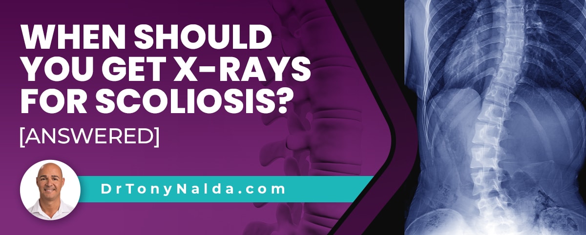
While some worry about radiation exposure, X-rays remain the safest and most reliable diagnostic tool, and scoliosis x-rays are integral to diagnosing, treating, and monitoring scoliosis. Continue reading to learn how scoliosis is diagnosed, treated, and monitored via x-ray.
Each case of scoliosis is unique, which is why the customization of treatment plans is so important. X-rays should be done as part of the diagnostic/assessment process, and throughout treatment to monitor how the spine is responding.
There is no better way to monitor what's happening in the entire spine than by x-ray image, so let's start by exploring the parameters that have to be met to reach a diagnosis of scoliosis.
Table of Contents
Diagnosing Scoliosis Via X-Ray
There are a number of spinal conditions that involve a loss of its healthy curves, but scoliosis has a number of condition-characteristics that set it apart from the rest.
And as current estimates have close to seven million people currently living with scoliosis in the United States alone, the condition is highly prevalent and warrants awareness.
Scoliosis involves the development of an unnatural lateral bending spinal curve, and the rotational element means the spine also twists, making scoliosis a 3-dimensional spinal condition.
As a 3-dimensional structural spinal condition, its underlying nature involves a structural abnormality within the spine itself, and this unnatural bend and twist in the vertebrae (bones of the spine) has to be assessed and further classified, and this process involves a series of comprehensive images taken by an x-ray machine, and measurements based on the x-ray images.
In addition to an unnatural bend and twist, a scoliotic spine has to have a minimum Cobb angle measurement of at least 10 degrees, and this measurement is taken during x-ray by drawing lines from the tops and bottoms of the curve's most-tilted vertebrae, at its apex; the resulting angle is expressed in degrees, and this measurement is also what condition severity is classified by:
- Mild scoliosis: Cobb angle measurement of between 10 and 25 degrees
- Moderate scoliosis: Cobb angle measurement of between 25 and 40 degrees
- Severe scoliosis: Cobb angle measurement of 40+ degrees
- Very-severe scoliosis: Cobb angle measurement of 80+ degrees
 When the spine's natural and healthy curves are in place, its vertebrae are aligned as they should be, but if an unnatural spinal curve develops, this means the spine is no longer aligned because some of its vertebrae have become unnaturally tilted, bending and twisting in unhealthy ways.
So x X-rays are needed to confirm a patient's Cobb angle, which is a key piece of information needed to reach an official diagnosis of scoliosis.
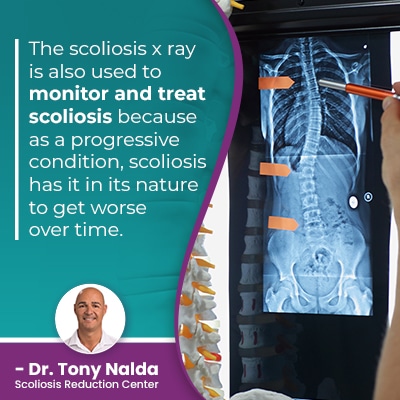
The scoliosis x-ray is also used to monitor and treat scoliosis because as a progressive condition, scoliosis has it in its nature to get worse over time.
Scoliosis Progression and X-Rays
As a progressive condition, most cases of scoliosis are guaranteed to get worse at some point, and that means the size of the unnatural spinal curve is increasing, as are the condition's uneven forces, and their effects.
Because scoliosis isn't a static condition, where it is at the time of diagnosis doesn't mean that's where it will stay, especially without the help of proactive treatment that works towards counteracting the condition's progressive nature.
In order to monitor what's truly happening in and around the spine, scoliosis x-rays are needed, and these are performed at various intervals, depending on each individual case.
Here at the Scoliosis Reduction Center, I order X-rays throughout treatment to see how the spine is responding as this information allows me to adjust treatment plans accordingly; part of my conservative chiropractic-centered treatment approach involves integrating multiple condition-specific forms of treatment so conditions can be impacted on every level.
Monitoring via x-ray allows me to see how the spine is responding to particular types of treatment so I can constantly adjust plans so they are further customized to address the specifics of each patient's scoliosis.
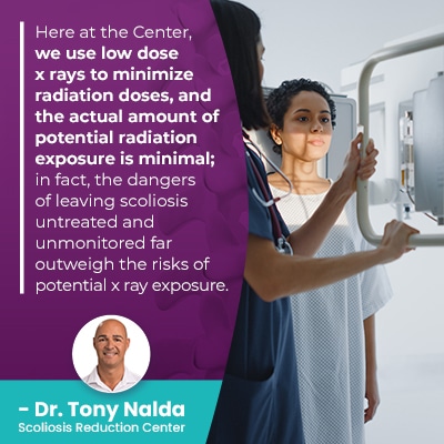
Here at the Center, we use low dose x-rays to minimize radiation doses, and the actual amount of potential radiation exposure is minimal; in fact, the dangers of leaving scoliosis untreated and unmonitored far outweighs the risks of potential x-ray exposure.
Growth and Progression
There are different types of scoliosis, but the most common type to affect both children and adults is idiopathic scoliosis, and the most prevalent type overall is adolescent idiopathic scoliosis (AIS), diagnosed between the ages of 10 and 18.
Idiopathic means not clearly associated with a single-known cause, and while we don't fully understand what triggers the initial onset of idiopathic scoliosis, we do know what triggers its progression: growth and development.
 Part of the focus of scoliosis treatment in children, and particularly adolescents who are in, or are entering into, the stage of puberty is monitoring scoliosis throughout growth to see how growth is affecting progression.
When treating adolescent idiopathic scoliosis (AIS), the goal is to achieve a significant curvature reduction and hold it there, despite the constant progressive trigger of growth, and the only way to monitor if an actual curvature reduction is occurring is to confirm a patient's Cobb angle measurement throughout treatment; x rays are needed to monitor how a patient's scoliosis is responding to growth/treatment.
As adolescents are at risk for rapid-phase progression because of the rapid and unpredictable growth spurts associated with puberty, I order X-rays more frequently than with my older patients, and while each case is unique, a typical case would start with x rays being performed every 3 months, and frequency is reduced alongside treatment success.
There are three main sections of the spine: the cervical spine (neck), thoracic spine (middle/upper back), and the lumbar spine (lower back).
X-ray images also tell us where along the spine the scoliosis has developed, and this helps us determine a condition's likely effects; in most cases, the area of the body located the closest to the affected spinal section is going to feel the majority of the condition's direct effects.
Patients and parents need to understand that accurate assessment of scoliosis is necessary not only to diagnosis and properly classify conditions, but also to monitor scoliosis for progression, and how the spine is responding to treatment; the information provided by x-ray imaging informs the crafting and customization of potentially-effective treatment plans.
The worst thing a patient can do is leave their scoliosis untreated because this means it's not being monitored properly, and this can mean allowing a scoliotic curve to progress unimpeded, and it's far more effective to proactively work towards preventing progression and increasing condition-effects, than it is to attempt to reverse those effects once they've developed.
Conclusion
When should you get x-rays for scoliosis is an excellent question to be asking because knowing the answer means understanding the condition's progressive nature.
Physicians need to monitor scoliosis through X-rays to see what's happening in and around the spine; a patient's Cobb angle is needed for diagnosing, assessing, and classifying scoliosis, and this is a measurement taken during x x-ray.
In addition to getting an X-ray done to confirm a diagnosis, X-rays are also needed to monitor for progression, particularly in child and adolescent patients who are at risk for rapid-phase progression
Most cases of scoliosis don't require surgery, but only if proactive treatment has been applied, and part of proactive treatment involves adjusting treatment plans accordingly in an effort to prevent progression and increasing condition-effects.
Here at the Center, my patients benefit from a modern conservative treatment approach that combines chiropractic care, physical therapy, corrective bracing, and rehabilitation, and in order to correct scoliosis, x-rays have to be performed so treatment plans can be fully customized; the more specific information about a patient's curvature type, location, and angle of trunk rotation, the more targeted treatment results can be.
Once skeletal maturity has been reached, progression tends to slow down, and while there is no single test to determine, with 100-percent accuracy, a patient's specific rate of progression, certain factors such as a patient's risser sign can be determined via x-ray, and this evaluation helps us determine how much growth a patient has yet to go through before reaching bone maturity.
A scoliosis specialist will be trained in how to comprehensively perform, measure, and interpret the results of a scoliosis x-ray, and while there are never treatment guarantees, monitoring scoliosis with how growth and treatment is affecting the spine via x-ray does increase the likelihood of treatment success.
X-ray imaging continues to be the gold standard in the diagnosing and assessment of a number of medical conditions; when performed safely by a professional x-ray technician, radiation exposure and radiation effects are minimal, and when compared with the dangers of allowing a progressive condition to progress unimpeded, the benefits far outweigh any potential risks.
Dr. Tony Nalda
DOCTOR OF CHIROPRACTIC
After receiving an undergraduate degree in psychology and his Doctorate of Chiropractic from Life University, Dr. Nalda settled in Celebration, Florida and proceeded to build one of Central Florida’s most successful chiropractic clinics.
His experience with patients suffering from scoliosis, and the confusion and frustration they faced, led him to seek a specialty in scoliosis care. In 2006 he completed his Intensive Care Certification from CLEAR Institute, a leading scoliosis educational and certification center.
About Dr. Tony Nalda
 Ready to explore scoliosis treatment? Contact Us Now


